
 FREE SHIPPING for online orders only over $75 with coupon code FRSHP - Limited Time Only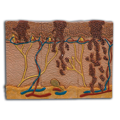
Skin Cancer Model
As low as $159.95
In the top view of this pathology model, the individual stages of externally visible skin changes are shown, allowing for an assessment according to the ABCDE criteria * The sides of the model show the various levels of invasion into the skin layers according to Clark (I-V) and the tumor thickness according to Breslow (in mm)
- 5 original color illustrations on the base show various types of malignant melanomas
- Shows the 6 different stages of a malignant melanoma on the front and back, enlarged 8 times: Healthy
- Malignant cells are found at the surface, within the epidermis
- Malignant cells fill the epidermis, a few invade the papillary layer
- Malignant cells fill the papillary layer
- Malignant cells invade the reticular layer
- Malignant cells have reached the subcutaneous fatty tissue, satellite cells approach a vein
- Supplied on a base
- 14x10x11.5 cm
This product features one of our many Healthcare Wellness and Anatomy designs.

Reviews
There are no reviews yet.